Orthopedic implants restore movement when bone, cartilage, or joints can no longer function normally. Their surfaces may remain inside the body for years. This makes material safety a clinical priority, not a marketing preference. The World Health Organization estimates that 1.71 billion people live with musculoskeletal conditions worldwide. As demand for joint reconstruction grows, implant reliability matters to patients, surgeons, and healthcare systems.
Why do orthopedic implants need to be biocompatible? The body identifies an implant as foreign, even when it is carefully engineered. Poor material selection may trigger inflammation, infection, allergic reactions, bone loss, or implant loosening. A painful hip or knee can turn a successful operation into another hospital visit. The FDA’s 2023 guidance on ISO 10993-1 recommends evaluating biological risks according to implant materials, contact tissues, and exposure duration. This approach supports evidence-based testing rather than assumptions.
Biocompatibility does not mean complete biological neutrality. No implant is perfect. Titanium, cobalt-chromium alloys, stainless steel, ceramics, and polymers each present different benefits and limitations. Wear particles can accumulate around moving components. Surface texture can also influence bone attachment and cellular behavior. The American Joint Replacement Registry and national registries, including the UK National Joint Registry, track revision patterns to evaluate long-term performance. These reports offer valuable real-world evidence, although registry data may not explain every patient outcome. Therefore, implant design requires laboratory testing, clinical judgment, manufacturing control, and long-term follow-up. Patient anatomy, activity level, surgical technique, and existing health conditions still matter. That detail is easy to overlook.
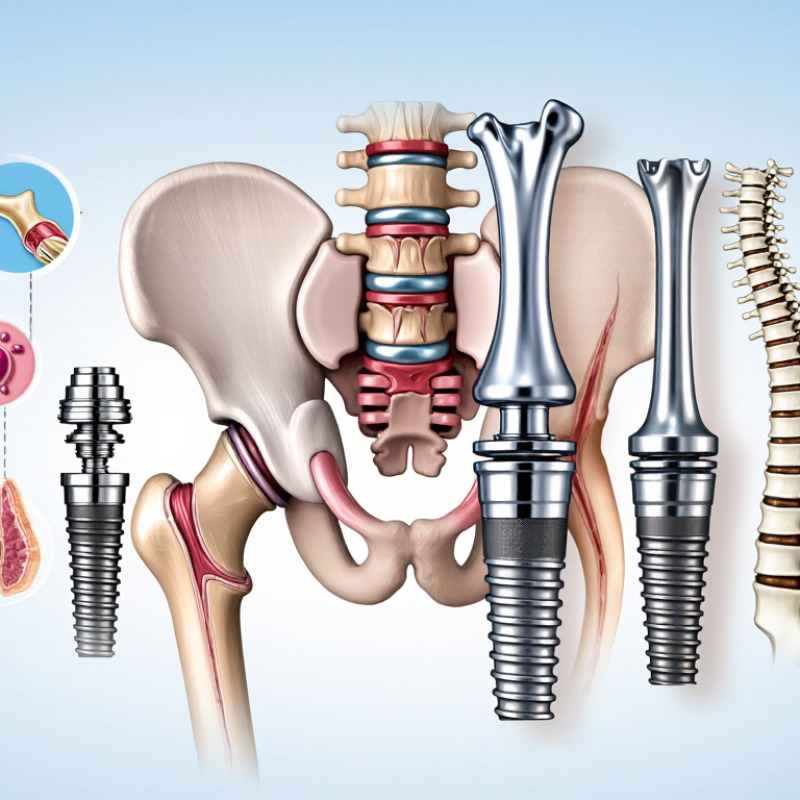
Biocompatibility means an orthopedic implant can work inside the body without causing harmful biological reactions. It is more than being “non-toxic.” Engineers examine corrosion, wear particles, inflammation, allergy, and tissue integration. A titanium component may appear stable, yet microscopic debris can irritate surrounding tissue over time. The FDA’s ISO 10993 framework evaluates biological safety through chemical, toxicological, and biological evidence. Testing cannot perfectly copy a moving knee, however. Real patients heal differently.
Clinical data adds necessary perspective. The UK National Joint Registry’s 21st Annual Report tracks more than three million primary hip and knee procedures. Its findings show revision risk varies with implant design, fixation method, age, and patient health. Biocompatibility therefore connects laboratory results with long-term outcomes. A material that performs well in a short study may behave differently after years of loading, moisture, and repeated movement. That uncertainty deserves attention.
Tips: Ask how the material was tested. Check evidence for wear, corrosion, and tissue response. Do not treat “medical-grade” as a complete safety answer. Surgeons should review implant history, patient allergies, bone quality, and activity level together. Patients also need realistic expectations. Even a well-tested implant can face infection, loosening, or unexpected inflammation. I once viewed biocompatibility as a simple pass-or-fail property; that was too narrow. It is better understood as an ongoing relationship between material, body, design, and clinical practice.
When an orthopedic implant enters the body, tissues do not treat it as empty space. Cells detect its surface, chemistry, stiffness, and tiny particles. Blood proteins attach within seconds, shaping later immune activity. Macrophages may gather around the implant and release signaling molecules. Some inflammation is expected. Persistent inflammation is not. That difference matters during healing.
Bone may grow tightly against a suitable surface, creating stable fixation. This process depends on implant design, bone quality, loading, and surface texture. Microscopic movement can interrupt new bone formation. Instead, fibrous tissue may develop around the device. The implant can then feel loose or painful. Material wear adds another concern. Repeated motion may produce microscopic debris, which nearby cells must process. Some particles remain quiet; others provoke stronger reactions. Metal sensitivity is uncommon, but clinicians still review allergies and previous reactions.
Biocompatibility is therefore not a simple label. It changes with time, anatomy, surgical technique, and patient health. Laboratory testing studies corrosion, wear, toxicity, and mechanical performance before clinical use. Surgeons also consider evidence, patient factors, and intended loading. Yet no test predicts every human response. That limitation deserves honesty. Even a well-studied material can behave differently in a smoker or patient with poor bone density. Infection can alter the tissue environment and threaten fixation. Careful follow-up helps distinguish normal healing from an adverse reaction.
Why Do Orthopedic Implants Need to Be Biocompatible?
Biological Risks of Incompatible Orthopedic Implants
An orthopedic implant stays close to bone, muscle, and blood for years. If its materials irritate the body, persistent inflammation may develop around the surgical site. Patients might notice swelling, warmth, tenderness, or reduced movement. Sometimes, the warning signs are subtle.
Incompatible materials can release microscopic particles through wear or corrosion. The immune system may react to this debris. Over time, bone loss can occur around the implant, weakening its fixation. This process, called osteolysis, may lead to loosening or revision surgery. Poor tissue integration creates another concern. The implant may appear stable at first, yet gradually lose support. Infection is also possible, although it does not always result from material incompatibility. These causes can overlap, which makes diagnosis difficult.
Tips: Discuss allergies, previous implant reactions, and chronic skin conditions before surgery. Ask how the implant’s material is tested for tissue compatibility. After surgery, report increasing pain, drainage, fever, or swelling promptly. Regular clinical reviews and imaging can reveal problems early. No implant is risk-free. Biological responses vary, and medical evidence is not perfect. A careful team should explain both the expected benefits and the remaining uncertainties.
Biocompatibility means more than avoiding immediate toxicity. An orthopedic implant must remain stable while the body responds to surgery, movement, and healing. In clinical practice, material selection considers corrosion resistance, fatigue strength, surface chemistry, and tissue response. These properties help reduce inflammation and support reliable integration with bone.
The implant’s elastic modulus also matters. If it is much stiffer than nearby bone, it may carry excessive load. Over time, surrounding bone can weaken from reduced mechanical stimulation. This effect is called stress shielding. A carefully designed material can transfer forces more naturally. Small changes matter here.
Surface texture influences how cells attach and grow. A controlled micro-texture may encourage bone integration, while excessive roughness can trap debris or complicate cleaning. The surface must also tolerate sterilization without losing its structure. Engineers test these features through mechanical cycles, chemical analysis, and biological evaluation. Still, laboratory results cannot represent every patient.
Body weight, activity level, allergies, bone quality, and surgical technique may change outcomes. No material is perfect. Even well-tested alloys and ceramics require careful monitoring after implantation. I think this is where material science needs humility: a strong result in testing is useful, but it is not a guarantee. Long-term evidence and patient follow-up remain essential for safe implant integration.
Why Do Orthopedic Implants Need to Be Biocompatible?
Implant biocompatibility means more than avoiding immediate toxicity. The device must work safely with bone, blood, muscle, and surrounding tissue. Evaluation usually begins with material screening and chemical analysis. Engineers examine corrosion, wear particles, surface residues, and possible allergic reactions. Laboratory tests may assess cell survival, inflammation, and tissue irritation. Standards such as ISO 10993 help organize this process, but they do not replace clinical judgment. Animal studies and long-term clinical evidence can reveal problems that short tests miss.
Tips: Review the whole implant system, not one material alone. Check coatings, cement, debris, sterilization, and packaging. Ask how testing reflects real movement, repeated loading, and aging. Small particles matter.
Improvement often starts with surface design. A smoother surface may reduce unwanted debris, while a carefully engineered texture can support bone attachment. Manufacturers may adjust alloy composition, polishing methods, coating thickness, or sterilization conditions. Each change needs verification because improving one property can weaken another. For example, a harder coating may resist wear but crack under repeated stress. Manufacturing controls are equally important. Tiny residues or uneven surfaces can change tissue responses. A clean laboratory result is encouraging, not final. Real patients vary in age, activity, immune response, and bone quality. That uncertainty deserves honest monitoring, careful follow-up, and continued reassessment after implantation.
| Biocompatibility Dimension | Why It Matters for Orthopedic Implants | Typical Evaluation Approach | Important Indicators | Common Improvement Measures |
|---|---|---|---|---|
| Cytotoxicity | Implants can release soluble substances or degradation products that may damage cells near the bone or soft-tissue interface. | Extract-based in vitro cell studies are commonly performed using cultured mammalian cells, with evaluation of cell viability and morphology. | Cell viability, membrane damage, cell morphology, and dose-dependent response. | Remove process residues, control alloy composition, reduce particulate contamination, and optimize surface treatments. |
| Sensitization and Irritation | Some chemical constituents or residues may trigger allergic contact reactions or local irritation after implantation. | Chemical characterization, toxicological assessment, and appropriately selected sensitization or irritation studies based on the device’s contact type and duration. | Potential sensitizing substances, inflammatory response, exposure level, and contact duration. | Use well-characterized materials, eliminate cleaning agents and residues, and apply validated manufacturing controls. |
| Systemic Toxicity | Wear debris, corrosion products, or leachable substances may enter circulation and affect organs away from the implant site. | Chemical characterization and toxicological risk assessment are combined with relevant acute, subacute, or chronic systemic studies when justified. | Chemical identity, concentration, exposure route, duration, and toxicological threshold. | Reduce leachable substances, improve corrosion resistance, control wear, and verify cleaning and sterilization processes. |
| Genotoxicity and Carcinogenicity Risk | Long-term exposure to certain degradation products or impurities may create genetic or tumor-related concerns. | A risk-based assessment considers material chemistry, degradation behavior, exposure, literature, and targeted genotoxicity testing when appropriate. | Mutagenic potential, impurity profile, degradation products, and cumulative exposure. | Select stable materials, limit impurities, characterize degradation products, and minimize uncontrolled surface degradation. |
| Local Tissue Response | Excessive inflammation, fibrosis, or foreign-body reaction can cause pain, loosening, delayed healing, or implant failure. | Histopathology, inflammatory marker analysis, imaging, and animal studies may be used when bench and clinical evidence are insufficient. | Inflammatory cell infiltration, fibrous capsule formation, necrosis, and tissue integration. | Control surface chemistry and roughness, reduce debris generation, and optimize implant geometry and fixation. |
| Hemocompatibility | Implants that contact blood during or after surgery may contribute to platelet activation, coagulation, or blood-cell damage. | Selected in vitro blood-interaction tests evaluate thrombosis, coagulation, platelet activation, or hemolysis when blood contact is clinically relevant. | Hemolysis, platelet adhesion, thrombin generation, coagulation time, and complement activation. | Improve surface cleanliness and finish, reduce sharp or irregular features, and select surface treatments suitable for the intended blood contact. |
| Corrosion and Ion Release | Body fluids can promote electrochemical corrosion, generating ions or particles that may cause local or systemic biological effects. | Electrochemical testing, immersion studies, ion-release analysis, and examination of the surface after simulated physiological exposure. | Open-circuit potential, corrosion current, mass loss, ion concentration, and surface damage. | Use corrosion-resistant materials, reduce galvanic couples, improve passivation, and control surface defects. |
| Wear and Particulate Debris | Particles generated at articulating or contacting surfaces may stimulate inflammation, macrophage activity, osteolysis, and implant loosening. | Tribological testing measures wear under representative load, motion, lubrication, and cycle conditions; particles are then characterized. | Wear rate, particle size distribution, particle morphology, composition, and total debris quantity. | Optimize material pairing, surface hardness, finish, alignment, lubrication conditions, and manufacturing precision. |
| Osseointegration | Stable bone attachment helps distribute load and reduces micromotion, fibrous encapsulation, and loosening. | Surface characterization, cell-attachment studies, mechanical fixation testing, imaging, and histological assessment may be used. | Bone-to-implant contact, pull-out strength, interfacial strength, cell attachment, and new bone formation. | Create controlled macro- and microtopography, improve surface cleanliness, and use validated coatings or chemical modifications. |
| Bacterial Adhesion and Infection Risk | An implant surface can provide a site for bacterial attachment and biofilm formation, making infection difficult to treat. | Surface microbiology studies, biofilm models, sterility testing, and clinical infection surveillance are selected according to the device and intended use. | Bacterial attachment, viable colony count, biofilm biomass, surface cleanliness, and sterility assurance. | Use validated sterilization, control surface contamination, optimize surface properties, and assess any antimicrobial strategy for safety and durability. |
| Degradation and Long-Term Stability | Material changes over time can alter mechanical performance, surface chemistry, debris generation, and biological response. | Accelerated aging, simulated body-fluid exposure, mechanical fatigue, chemical analysis, and post-test surface examination. | Mass change, mechanical-property retention, surface alteration, degradation-product profile, and fatigue life. | Select stable materials, validate coatings, limit residual stresses, and verify performance after aging and sterilization. |
| Sterilization and Process Residues | Sterilization can change material properties or leave residues that affect tissue compatibility and implant performance. | Sterilization validation, residual analysis, material compatibility testing, and functional testing after sterilization. | Sterility assurance, residual chemicals, endotoxin level, discoloration, dimensional change, and mechanical-property retention. | Validate cleaning and sterilization parameters, improve packaging, control endotoxin, and reassess the device after repeated or final sterilization exposure. |
| Mechanical–Biological Interaction | Excessive stress, micromotion, mismatch in stiffness, or fatigue damage can indirectly produce harmful tissue responses. | Fatigue, wear, fixation, finite-element, and mechanical-performance testing are interpreted together with biological and clinical evidence. | Micromotion, stiffness, fatigue strength, load distribution, fixation stability, and fracture risk. | Optimize geometry and material properties, improve fixation design, reduce stress concentrations, and verify performance under realistic loading. |
It means the implant can function inside the body without harmful biological reactions. It must interact safely with bone, blood, muscle, and nearby tissue. It is more than being non-toxic.
Corrosion resistance, fatigue strength, surface chemistry, and tissue response are important. The material should remain stable during movement, moisture exposure, and healing. Small changes matter.
An implant much stiffer than bone may carry too much load. This can reduce natural stimulation and weaken nearby bone over time. This effect is called stress shielding.
A controlled texture may help bone cells attach and grow. Excessive roughness can trap debris or make cleaning harder. A coating may resist wear, but it can crack under repeated stress.
Testing may examine chemical residues, corrosion, wear particles, cell survival, inflammation, and tissue irritation. Mechanical cycles can imitate repeated loading. Laboratory tests are useful, but they cannot copy every moving knee.
Ask how the material was tested and whether long-term clinical evidence exists. Check evidence about wear, corrosion, allergies, and tissue response. “Medical-grade” is not a complete safety answer.
Yes. Infection, loosening, inflammation, or unexpected tissue reactions can still occur. Patients heal differently. A strong test result is encouraging, not a guarantee.
Age, body weight, activity level, allergies, bone quality, immune response, and surgical technique may affect results. A highly active person may place greater repeated stress on the implant.
Engineers may adjust alloy composition, polishing, coating thickness, or sterilization methods. Manufacturing must control tiny residues and uneven surfaces. I once saw biocompatibility as pass or fail. That was too narrow. Continued follow-up remains necessary.
Why do orthopedic implants need to be biocompatible? Biocompatibility is essential because an implant must function safely inside the body without causing harmful reactions. A biocompatible orthopedic implant is designed to work with surrounding bone, muscle, and tissue while supporting healing, stability, and long-term mobility. When the body encounters an implant, the immune system may respond to its material, surface, or wear particles. A suitable material should therefore minimize inflammation, toxicity, allergic reactions, infection risks, and unwanted tissue formation.
Safe implant integration depends on properties such as strength, corrosion resistance, durability, appropriate stiffness, and a surface that encourages healthy bone attachment. Biocompatibility is evaluated through laboratory analysis, material testing, mechanical assessment, and carefully controlled biological studies. Researchers and manufacturers can improve implant safety by refining material composition, surface structure, manufacturing quality, and sterilization methods. Together, these considerations help ensure that orthopedic implants remain stable, functional, and well tolerated throughout their intended service life.

Medev Medical